| The Determination of RBE of Soft X-rays | |||
|---|---|---|---|
| A. Panteleeva, K. Brankovic1, W. Dörr1, W. Enghardt, J. Pawelke, D. Slonina2 | |||
|
The superconducting electron linear accelerator ELBE, at present
under construction at Forschungszentrum Rossendorf, is going to be
used to produce various types of secondary radiation, among which
are X-rays in the energy range 100 eV - 100 keV. At the first
stage, monochromatic X-rays in the energy range 10 keV - 50 keV
will be obtained by means of planar channeling of high-energy
electrons in a diamond crystal. Such an unconventional X-ray source
is rather compact, tuneable and allows the possibility of delivering
a beam in picosecond pulses [1]. The relative biological
effectiveness (RBE) of the X-rays in this energy range will be
determined by studies of cell survival and cytogenetic damage.
Precise RBE values are required
for risk assessment in diagnostic radiology, such as mammography,
and radiotherapy with soft X-rays (brachytherapy). It has been previously
shown that
soft X-rays are more effective in cell killing [2] and chromosome
abberations induction [3]. In preparation of radiobiological studies
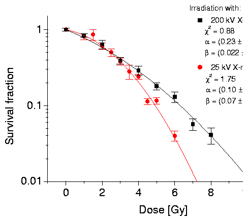
at ELBE, experiments were performed with a 25 kV soft X-ray tube
and a 200 kV reference X-ray tube at TU Dresden. The cell line
NIH/3T3 mouse fibroblasts was chosen because it is widely used,
easy to handle, and contact-inhibited. The cells were cultured in
DMEM supplemented with 10 % bovine serum, 10 mM HEPES buffer,
1 mM Sodium Pyruvate, 1 % non-essential aminoacids,
100 U/ml penicillin and 100 mg/ml streptomycin (all from
Biochrom Seromed) at 37oC in air containing 5 % CO2.
Exponentially growing cells were irradiated in 25 cm2
polystyrene flasks at dose rates 1.22 Gy/min
(200 kV, 20 mA, 0.3 mm Al filter) and 1.67 Gy/min
(25 kV, 20 mA, 0.5 mm Cu filter). After irradiation the cells were
harvested, counted and seeded at appropriate densities (6-8
replicates per dose point). After 12 days, colonies having at least
50 cells were scored as offspring from 1 surviving cell.

Fig. 1 Clonogenic survival of NIH/3T3 cells after 25 kV and 200 kV X-ray tube irradiation.
1 Dresden University of Technology, Germany
References
[1] W. Enghardt et. al., Acta Physica Polonica, Vol. 30 (1999), No. 5, pp. 1639-1645
|