Motivation
For operation of an FEL the overlap of the electron beam and the
optical beam inside an undulator must be maximized. Both of
them must be located in the magnetic axis of the undulator.
To realize this one must be able to measure the electron beam
position and profile inside the undulator with an accuracy of
about 100 mm. These requirements are connected to the size of the
electron beam and the optical mode in the undulator.
Scheme of the diagnostic system
The proposed diagnostics is shown on Fig 1. The system works
in the following way: a moveable screen travels in the vacuum
chamber which is located in the undulator. The travel length
of the screen in the case of the undulator U27 is the length of
the undulator (2 × 98 cm) plus a gap between the two parts of the
undulator ( ~ 30 cm). In the case of undulator U50 the travel
length is approximately the length of the undulator ( ~ 225 cm).
That means the travel length of the screen is approximately the
same for both undulators.
When the electron beam hits the YAG screen radiation with
maximum intensity at 550 nm is produced.
Downstream of the undulator there is a dipole magnet to
bend the electron beam. The distance between the undulator and the
dipole is approximately 140 cm. An insertable mirror is located
behind the dipole. The mirror is used to deflect the YAG
radiation from the vacuum chamber though a vacuum window.
Near the vacuum window a lens is placed for imaging the
screen to a CCD chip. Since the screen is moveable and we
need to keep the optical path length between the screen
and the lens constant we have to insert an element which
has a variable optical path length. We can use a moveable roof
mirror plus two mirrors 2 and 3, see Fig 1. The travel
length of the roof mirror is half of the screen travel length.
The mirror 4 and a very sensitive cooled CCD camera are located
on the floor. The camera has to be shielded by a lead housing.
Choice of the screen material
As also hard X-rays will be produced, when the electron beam
strikes the screen, the undulator magnets may loose magnetic
strength in this high radiation environment; thus
the screen should be as thin as possible. From this
point of view an OTR screen would be preferable, since it
is possible to make such screen from aluminium foil as thin as
10 mm. But in diagnostic mode with average current
about 1 mA at an electron beam energy of
15 MeV ¸ 40 MeV the radiation level in our collection
solid angle is too small. Thus using an OTR is not sufficient
for this diagnostic. The YAG crystal is a good alternative to
the OTR screen in this case. Direct comparisons of the
effective conversion efficiency, spatial resolution, and
time response of the screens have been performed [1,2].
The light intensity of the YAG crystal is about 1000 times
larger than with the OTR screen. A beam size growth has been
observed for the YAG data relative to the OTR data above
average current 60 mA. The fluorescence time of a the crystal
has been measured [2]. The light intensity decay time was
measured to be about 80 ns. YAG crystals with thickness
0.1 mm are commercially available [3].
The cooled CCD ST-237 [4] was chosen for the diagnostic,
since it is sensitive enough and still not too expensive.
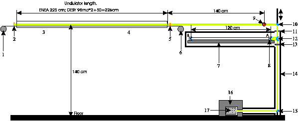
Fig. 1 Scheme of the diagnostics.
1 - stepmotor 1, 2 - screen in position A, 3 - first part of
the undulator U27, 4 - second moveable part of the undulator,
5 - screen in position B, 6 - stepmotor 2, 7 - linear motor stage
with travel length 1.3 m, 8 - roof mirror on position A, 9 - position
of the dipole, 10 - insertable mirror, 11 - window, 12 - mirrors,
13 - lens, 14 - light shielding, 15 - mirror, 16 - lead shielding,
17 - CCD camera.
References
|
[1]
|
A.H. Lumpkin, Nucl. Instr. and Meth. A 429 (1999)
336-340
|
|
[2]
|
W.S. Graves, E.D. Johnson, A high resolution electron
beam profile monitor,
|
|
|
Proc. of Particle Accelerator Conf. 1997, Vancouver, Canada, Vol. 2, p. 1993
|
[3]
|
http://www.crytur.cz/
|
|
[4]
|
http://www.sbig.com/sbwhtmls/online.htm
|
IKH
05/21/01
© P. Evtushenko
|