| Possible Effect of the Carbon Ion Beam Microstructure on In-Beam PET Measurements at GSI Darmstadt B | ||||||
|---|---|---|---|---|---|---|
| K. Parodi, W. Enghardt, H. Eickhoff1, T. Haberer1, P. Moritz1, P. Forck1, A. Peters1 | ||||||
|
The data acquired by the positron camera during the beam spills delivered by the heavy ion synchrotron SIS at GSI Darmstadt are affected by high noise level and are useless for the image reconstruction. A reason could be a wrong correction of random coincidences. The standard method of random correction, identifying random coincidences with delayed ones (delay time of 128 ns and coincidence window 12 ns wide), properly works in nuclear medicine, where the random background seen by the scanner is rather constant in time. In the therapy case, a background correlated in time with the beam microstructure on a time scale comparable to the delay time could lead to an underestimation of random coincidences and hence to the observed increase of noise. 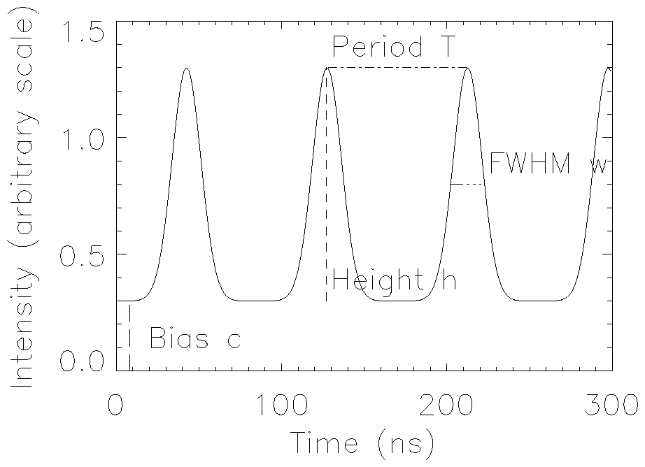
Fig. 1. Model of the background radiation reaching the scanner during the beam spill.
In Fig. 1 we sketch the background model used in our simulation approach for reproducing the experimental random (i.e. not related to b+-activity) prompt and delayed coincidence rates acquired by the camera in spill. The periodical repetition of the bunch Gaussian shape represents the background radiation directly following the beam appearance, whereas the bias c introduces a delayed g-ray
component, due to isomeric de-excitations or radioisotope decay with half-lives longer than several ns.
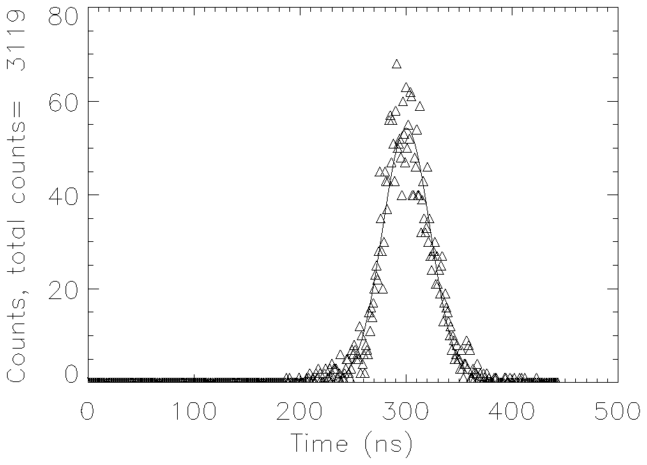
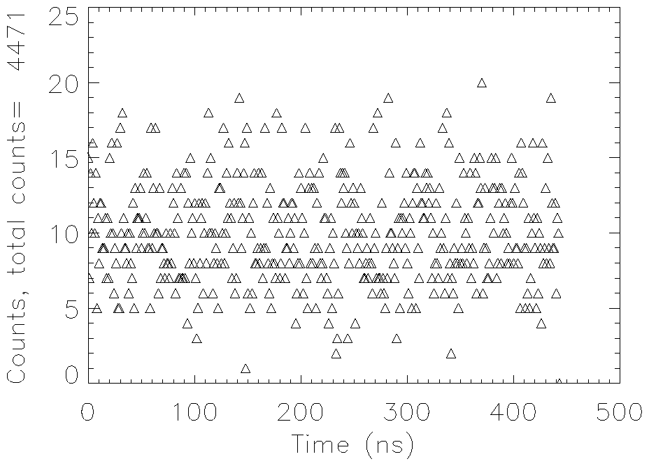
Fig. 2. Left: example of beam microstructure at Uft = 2 kV. A Gaussian fit is superimposed onto the data. Right: example of not correlated time distribution measured at Uft = 0 kV. 1 Gesellschaft für Schwerionenforschung Darmstadt
|