| TSEE Dosimetry of Soft X-rays | ||||||||||||||||||||||||||||||||||||||||
|---|---|---|---|---|---|---|---|---|---|---|---|---|---|---|---|---|---|---|---|---|---|---|---|---|---|---|---|---|---|---|---|---|---|---|---|---|---|---|---|---|
| J. Pawelke, A. Panteleeva | ||||||||||||||||||||||||||||||||||||||||
|
The measurement of RBE by cell irradiation requires precise
determination of dose, delivered to the cell target.
Absolute dose measurements are usually done by an air-filled
ionisation chamber. However, in the case of low energy
X-rays (below 10 keV) it does not sample the cell target because of the
high attenuation. Detection based on thermally stimulated exoelectron
emission (TSEE) allows to sample the depth dose distribution in a cell
monolayer ( » 5 mm thick),
because the thickness of the sensitive detector layer is of the order of
~ 10 nm [1]. A TSEE prototype system
(Dr. Holzapfel Messgerätelabor, Teltow)
consisting of 3 types of thin-film BeO (100 nm) detectors and
a counter based on a spherical anode
Geiger-Müller methane gas-flow counter was tested.
The irradiation was performed with a
high-activity beta-source (90Sr),
a calibrated gamma-source (137Cs) and a soft
X-ray source (55Fe).
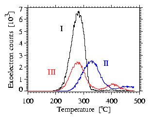
Highest response as well as stability were shown by detector type I
(fig. 1, table 1, same results were obtained with 55Fe source),
which proves that the 200 nm Au layer
contributes for better mechanical attachment of the BeO layer
as well as for electrical stability.
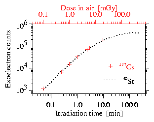
The saturation in the dose response
(fig. 2) due to the limited pulse resolution of the gas-flow counter
results in an upper detection limit of about 0.1 Gy and is far from
the requirements of the cell survival studies ( £ 10 Gy).
Therefore it cannot be used as online monitoring system.
In order to determine the dose delivered to cells,
measurements must be done in the presence of cell culture medium.
Irradiations were made after wetting the surface and showed
no change in the detector response due to any tribo-effect [2].
As expected detector response after irradiation of detectors covered with
about 0.5 mm propanol with low energy X-rays from 55Fe source
and high energy photons from 22Na source was reduced
only due to attenuation in the liquid layer.
However, the dependence of the detector
response on photon energy and dose rate must still be studied.
Table 1 Main detector properties.
a) average error of the mean for a series of
8 measurements with 90Sr
b) 137Cs, dose in air References
[1] J.S. Durham et. al., Radiation Protection Dosimetry,
Vol. 39(1991), No 1/3, pp. 67-70
|

