Magnetometry
1. Ferromagnetic Resonance - FMR
2. Low temperature Ferromagnetic Resonance
3. Microreosnator Ferromagnetic Resonance
4. Brillouin Light Scattering Microscope (BLS)
5. Time-Resolved Magneto-Optical Kerr Effect
6. Magneto-Optical Kerr Effect - MOKE
7. Kerr Microscopy
8. Probe Station for Nanostructures
9. Frequency-resolved MOKE for optically detected FMR
10. Atomic & Magnetic for Microscope
11. Atomic & Magnetic for Microscope
12. Electric Prober for Phase Change Materials
13. Vibrating sample magnetometer - VSM
| Bild |
Beschreibung |
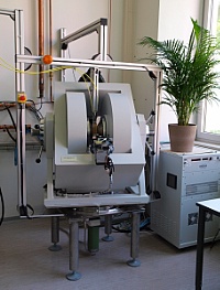 |
1. Ferromagnetic Resonance - FMR
- in-house development
- Agilent E8364B Vector Network Analyzer:
Frequency range 0.05 - 50 GHz
- Bruker Electromagnet: max. 2.2 T
- polar and azimuthal sample rotation
- Helmholtz magnet: max 0.1 T
Responsible: K. Lenz
top of page
|
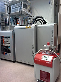 |
2. Temperature variable Ferromagnetic Resonance
- Attocube Attodry 1000 closed cycle cryostat
- 5 T split-coil magnet
- temperature range 4 - 300 K
- Agilent N5225A Vector Network Analyzer:
Frequency range 0.05 - 50 GHz
- full polar and azimuthal sample rotation
- SMU for resistancd and magnetoresistance measurements
Responsible: K. Lenz
top of page
|
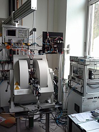 |
3. Microresonator Ferromagnetic Resonance
- in-house development
- microwave bridge
Frequency range 4-40 GHz
- Bruker Electromagnet: max. 2.2 T
- azimuthal sample rotation
- field modulation/Lock-in detection
Responsible: K. Lenz
top of page
|
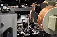 |
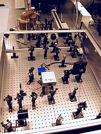
4. Brillouin-Lichtstreumikroskop 1+2
- in-house development
- BLS1: Phase- and time resolved
BLS2: time resolved only
- Tandem Fabry-Perot interferometer TFP-2 HC by JRS Scientific Instruments
- Sample positioning by XM Ultra-Precision Linear Motor Stages von Newport
- 50x and 100x objectives
- Magnet field up to 0.9 T
- Time resolution: down to 30ps using TimeTagger 20 from Swabian Instruments
- GSG Picoprobes for microwave input
- RF excitation up to 40 GHz, 30 dBm max.
- DC excitation by SMU
- arbitrary and pulse pattern generator
Responsible: H. Schultheiss
top of page/a>
|
 |
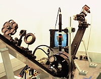
5. Time-Resolved Magneto-Optical Kerr Effect
- in-house development
- Delay time up to 5 ns
- Feld: max. 1 T
- Femtolasers XL500
- Wavelength: 800 nm
- Pulse-Length 40 fs
- Repetition rate: 5 MHz
- Output Power: 2.6 W
- Energy: 500 nJ/pulse
Responsible: H. Schultheiß
top of page
|
 |
6. Magneto-Optical Kerr Effect - MOKE
- in-house development
- longitudinal and transversal Kerr effect
- Field (in-plane): max. 400 Oe (Helmholtz coils)
- x, y, f scanning capabilities
- Temperature range: RT – 600 K
Responsible: H. Schultheiß
top of page
|
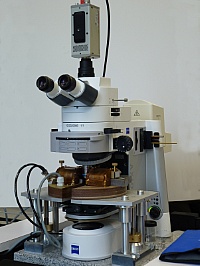 |
7. Kerr-Microscopy
- Manufacturer: Evico-Magnetics
- Microscope: Zeiss Axio Imager.D1m
- Light Source: two high-power LEDs (blue+red)
- Bipolar and quadrupol magnet
- twin-color system for quantitative Kerr analysis
- integrated AMR measurement
Responsible: H. Schultheiß
top of page
|
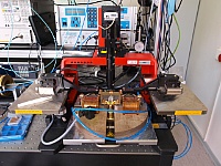 |
8. Probe Station for Nanostructures
- Süss MicroTech PM5 Wafer Prober
- Süss Z-Probes: GSG 0-50 GHz, 150µm Pitch
- Evico Magnetics electromagnet: Bmax= 0.6 T
- Optem CCD-Microscope
- Agilent MXA spectrum analyzer
- Picosecond PulseLab pulser
- Tektronix DPO72004 20GHz real-time oscilloscope
- Keithley source meter
Responsible: K. Lenz
top of page
|
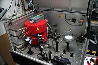 |
9. Frequency-resolved MOKE
- optically detected FMR (5 µm spatial resolution)
- Magnetic field up to 1.4 T
- Kepco power supply
- Frequency range up to 35 GHz
Responsible: K. Lenz
top of page
|
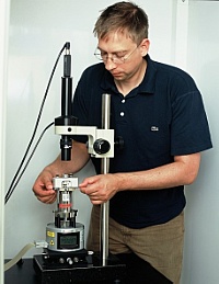 |
10. Atomic and Magnetic Force Microscope (AFM/MFM)
- Veeco/DI Multimode
- sample size: 10 x 10 mm²
- scan area max. 100x100 µm²
- scan area 15x15 µm²
- magnetic field 0 - 40 mT
- resolution:
- Scanning Spreading Resistance Microscopy (SSRM) option
Responsible: K. Potzger
top of page
|
 |
11. Atomic Force Microscope (AFM/MFM)
- Bruker Icon Dimension
- sample size: up to 20 x 20 cm²
- scan area max. 100 x 100 µm²
- AFM: ScanAsyst mode, tapping mode, contact mode
- MFM: lifted mode
- resolution: ~1nm
Responsible: K. Potzger
top of page
|
| |
12. Electric prober for phase change materials
- temperature and magnetic field dependence of resistance
- sample size: 10 x 10 mm²
- temperature RT - 450 K
- field: 1 kOe in-plane
- base pressure < 1e-6 mbar
Responsible: K. Potzger
top of page
|
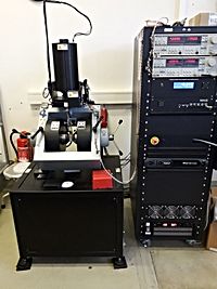 |
13. Vibrating Sample Magnetometer - VSM
- magnetic field max. ±1.8 T
- sensitivity: 5x10⁻⁶ emu (0.8x10⁻⁶ rms noise)
- angular range 360°
- vector coil option
- room temperature
responsible: R. Salikhov
|

