X-ray Equipment
Objectives
Ionising radiation such as X-rays is well suited for the investigation of multiphase flows since it penetrates all phases (gas, fluid, solid) well and is essentially linearily attenuated. Further, tomographic techniques give the opportunity to determine the volume density distribution of an object noninvasively. Beside two-dimensional tomography in an object’s plane an arrangement of X-ray source and 2D X-ray image detector can be used in combination with special cone-beam image reconstruction algorithms to reconstruct three-dimensional density distributions. In the frame of research on two- and multiphase flows we use radiographic and tomographic techniques for the investigation of gas, particle and phase distributions in flow channels and chemical reaction vessels.
The X-ray Measurement System
The department possesses a modern X-ray facility for radiography and tomography.
Panorama view of the X-ray facility with array detector, object rotation table,
and the two X-ray tubes.
The system consists of two medical X-ray tubes, a 2D X-ray detector array, a 64 element linear X-ray detector with high sampling frequency, and a rotational table for tomography of small objects. The detailed technical parameters of the system are given below. Depending on the type of investigation we can either use the integrating 2D detector array or the fast sampling linear detector in combination with either a continuous-wave X-ray radiator or a pulsed X-ray source. Thus, for short time exposures with down to 300 µs exposure time the PHILIPS ROTALIX SRO 22 50 source and for standard exposures the DUNLEE/PHILIPS PXD 1429 can be used. The data acquisition for both detectors is controlled by a measurement PC. The X-ray system is completely enclosed in a shielding tunnel that is closed during measurements. This guarantees sufficient radiation protection for the operating personnel. Since the arrangement of the different measurement components can be freely chosen for a given experiment we have a high degree of versatility for a number of scientific problems.
Data Acquistion and Processing Software
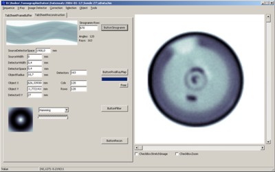
For computed tomography
we have developed a special software that controls the data acquisition
process in synchronization with the trigger for the X-ray source, and
further comprises an experimental version of different image reconstruction
algorithms (filtered backprojection, cone-beam reconstruction, iterative
image reconstruction). |
 |
|
| tomography software: example for tomographic imaging of a needle probe sensor. |
System Specification:
X-ray radiator
Radiator 1:
| x-ray generator | EDITOR MP601 |
| x-ray radiator | DUNLEE/PHILIPS PXD 1429 CS |
| focal spot size | 0.6 mm, 1.2 mm |
| maximum power for single exposures | 63 kW |
| acceleration voltage (single exposures) | 40...150 kV |
| maximum electron current (single exposures) | 10...630 mA |
| exposure time (single exposures) | 2...6300 ms |
| acceleration voltage (transillumination mode) | 40...125 kV |
| electron current (transillumination mode) | 1 mA |
| exposure time (transillumination mode) | user-defined |
Radiator 2 (pulsed radiator):
| pulse generator | RIG 150 |
| radiator | PHILIPS ROTALIX SRO 2250 |
| focal spot size | 0.6 mm; 1.2 mm |
| maximum pulse power | 56 kW |
| maximum pulse frequency | 300 Hz |
| pulse duration | ≥ 300µs |
X-ray Detectors:
X-ray Detector Array Perkin-Elmer RID 1640
| active array | a-Si:H, LANEX®-plane scintillator |
| resolution | 1024 x 1024 pixel |
| active pixel area | 400 µm x 400 µm |
| overall active area | 409.6 mm x 409.6 mm |
| resolution depth | 16 bit |
| maximum sampling rate | 3 frames/s |
X-ray Detector Line Array
| resolution | 64 pixel |
| active pixel area | 1.5 mm x 1.5 mm |
| length | 119 mm |
| resolution depth | 12 bit |
| maximum sampling rate | 100.000 kHz per pixel |
Measures of the shielding tunnel:
| height | 1.80 m |
| width | 1.10 m |
| depth | 3.20 m |

